


What are the key parts of a microscope, and how do they function? Our guide explores each microscope part to help you understand the mechanisms that bring tiny details into clear view. Without oversimplifying or overwhelming, we equip you with the knowledge to navigate the complex anatomy of microscopes.
Key Takeaways on Microscope Part
- Microscope parts come in three main types—light, electron, and fluorescent—each with its own set of specific applications, magnifications, and specialized components.
- Essential parts of a microscope include the eyepiece, objective lenses, light source, and focus knobs, and these function collectively to magnify and clarify specimens.
- Proper microscope maintenance, which includes regular cleaning and careful storage, is crucial for preserving image quality and extending the lifespan of the instrument. When purchasing, the choice of materials, construction quality, and selecting a reputable dealer are key considerations.
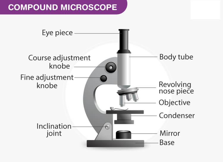
Understanding Compound Microscope Types
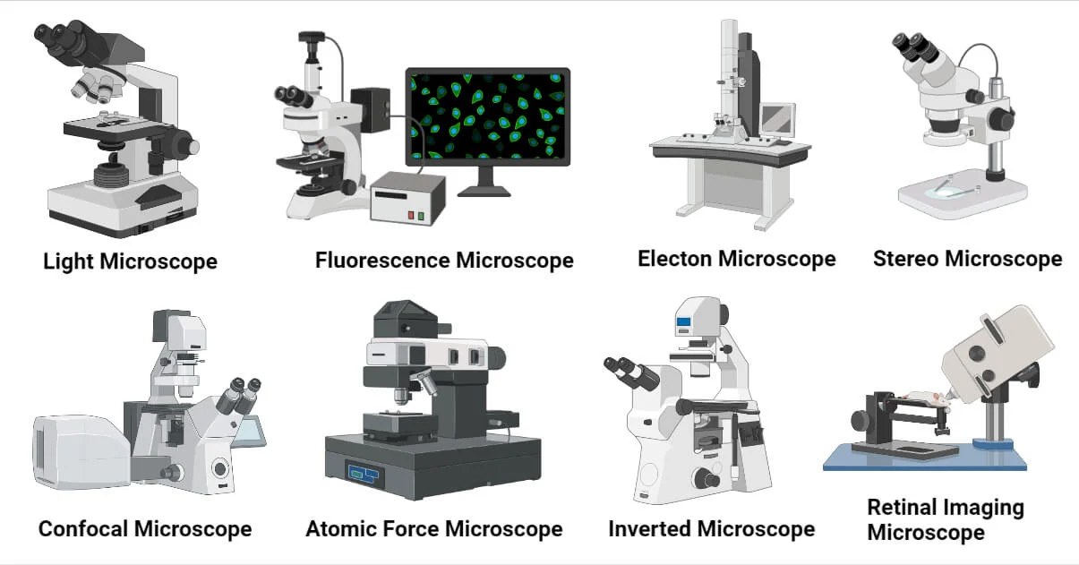

In the world of microscopy, there are three main types of microscopes, each with unique features: light, electron, and fluorescent microscopes. A stereo microscope, often used for dissection and detailed examination of larger specimens, adds another dimension to the applications of microscopy. A key player in high-power microscopy is the compound microscope, known for its multiple lenses, adjustment knobs, and applications in fields such as pathological and forensic labs, as well as in educational settings. These devices offer a glimpse into the intricate details of the world around us, from the tiniest microorganisms to the complex structures of cells.
Recognizing these types is a prerequisite to grasping the intriguing realm of microscopy.
Light Microscopes
A light microscope, often found in biology laboratories, uses visible light and a system of lenses to magnify tiny objects. This instrument’s beauty lies in its simplicity and accessibility, making it a popular choice for research and education. Light microscopes, including the binocular microscope, are versatile, with types ranging from:
- bright field
- phase contrast
- dark field
- fluorescence
Each type of stereo microscope is designed for specific applications. Whether you are observing the cells in an onion skin or examining bacteria in a yogurt sample, a light microscope provides a window into the unseen world.
While simple in design, light microscopes boast a sophisticated mechanism. The eyepiece tube, a vital component, connects the eyepiece lens to the objective lenses, forming an integral part of the light path. The eyepiece, in turn, magnifies the image produced by the objective lenses, allowing the observer to view the specimen in greater detail.
Electron Microscopes
Stepping outside the boundaries of light microscopy introduces us to electron microscopes. These high-tech devices use a concentrated beam of electrons, rather than light, to generate images. This enables them to provide a broader magnification range and superior resolution compared to their light-based counterparts. Imagine being able to discern structures as tiny as atoms – that’s the power of an electron microscope.
However, not all electron microscopes are made the same. There are two primary types of electron microscopes: the transmission electron microscope (TEM) and the scanning electron microscope (SEM). These powerful devices can achieve magnification levels as high as 10,000,000x and resolutions of up to 50 picometers (0.05 nanometers). From exploring the surface of a nanomaterial to peering inside a virus, electron microscopes open up a world of possibilities.
Fluorescent Microscopes
Fluorescent microscopes introduce yet another distinctive method of inspecting specimens. These optical devices use fluorescence to generate an image, creating vivid, multi-colored visuals that can highlight specific structures within a cell or tissue. The key to this process lies in the use of:
- a light source
- an excitation filter
- a dichroic mirror
- an objective lens
- a sample stage
These components work together to create an image that’s not just visually stunning, but also packed with valuable data.
So, where is the typical setting for the use of fluorescent microscopes? These specialized tools are commonly employed in disciplines such as biology, biomedical sciences, and materials science, where they can help visualize structures in life science specimens or assist in the visualization of latent fingerprints in forensic science. With the ability to examine a diverse array of fluorescent substances present in live or fixed organisms, tissues, or cells, fluorescent microscopes offer an illuminating perspective on the microscopic world.
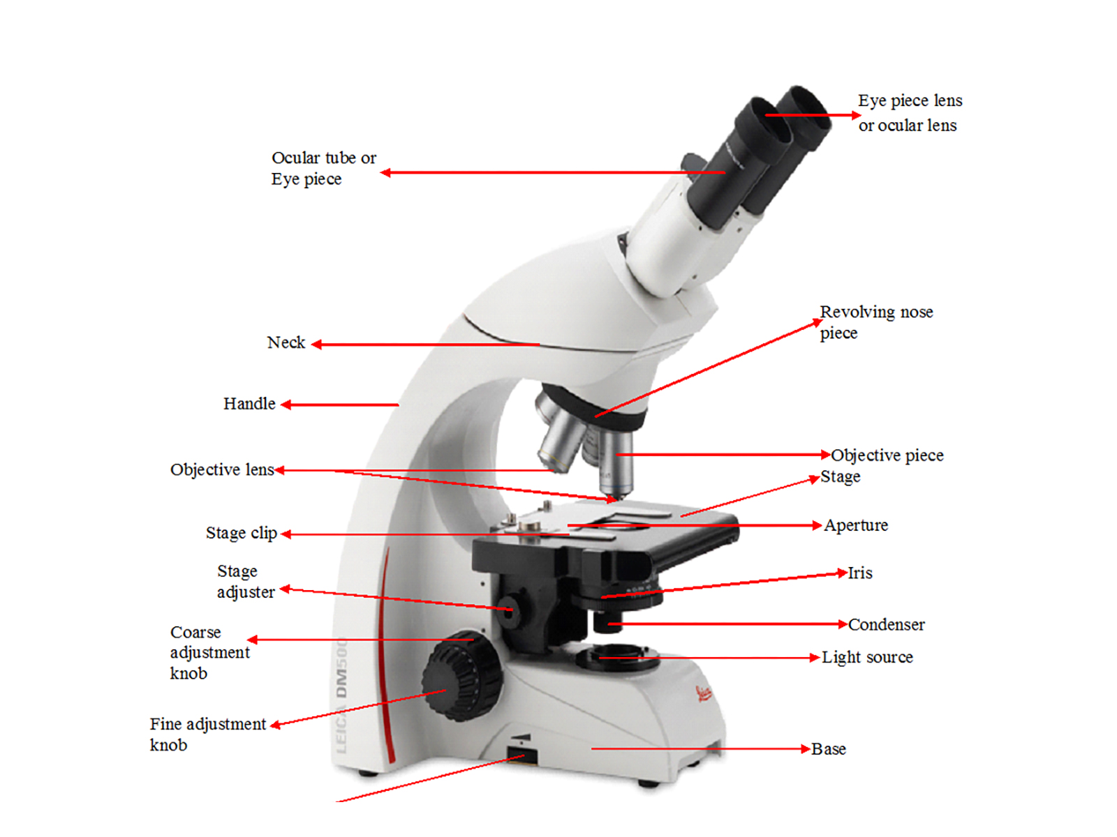
Key Components of a Microscope

Regardless of their type, all microscopes share some fundamental parts of a microscope central to their operation. These include:
- Eyepiece
- Objective lenses
- Light source
- Focus knobs
A compound microscope, for instance, incorporates these key components along with additional adjustment knobs, making it essential for high-power microscopy in fields such as pathological and forensic labs, as well as in educational settings.
Additionally, the iris diaphragm is a crucial component that controls the amount of light reaching the specimen, enhancing image clarity and contrast.
These elements, each with its specific role, work in harmony to make the invisible visible, unveiling the secrets of the microscopic world.
Eyepiece
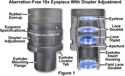

The eyepiece, or the ocular lens, holds a critical function in the microscope. It’s where we place our eye to view the magnified specimen, but it does more than provide a viewing portal. The eyepiece works in conjunction with the objectives to amplify the intermediate image, enabling us to see the specimen in greater detail.
Microscopes can have different types of eyepieces. The most common types have barrel diameters of 1.25 inches or 0.95 inches, which are also frequently used in telescope eyepieces. The quality of an eyepiece is influenced by the arrangement and curvature of its lens elements, which determine its total magnification power and field of view.
Objective Lenses

Objective lenses in a microscope:
- Capture light from the specimen
- Concentrate the light to generate a magnified image
- Interchangeable and mounted on a revolving nosepiece, allowing for easy switching between different magnification levels.
It is available in a range of magnification powers, with the most prevalent ones being:
- 4x
- 10x
- 40x
- 100x
These lenses are crafted from advanced glass formulations, such as fluorspar or newer synthetic materials, which enhance image quality and clarity.
Light Source
A light source to brighten up the specimen is indispensable for every microscope. This light source, often a bulb or an LED, sends transmitted light through a condenser, which focuses the light onto the specimen. Adjusting the light source allows us to control the intensity and focus of the light passing the illumination, thereby improving the clarity and contrast of the specimen.
The type of light source used can vary for most light microscopes, with some microscopes using:
- Incandescent lamps
- Halogen lamps
- Arc lamps
- LEDs
Adjusting the light source allows us to control the light intensity, and focus of the illumination, thereby improving the clarity and contrast of the specimen.
Focus Knobs
Focus knobs are crucial for achieving a sharp focus on the specimen, including the condenser focus knob. These knobs, which include both coarse focus and fine adjustment knobs, allow for precise tuning to enhance the clarity of the specimen being investigated.
The coarse adjustment knob facilitates rapid initial focusing. The fine adjustment knob enables finer tuning, especially for high-power lenses and under high magnification. Typically crafted from plastic or metal, these knobs offer reliable adjustment mechanisms.
Additional Microscope Parts and Their Functions
Apart from the principal parts of a microscope, a microscope head also encompasses other vital parts that add to its functionality. These include:
- The stage
- The condenser
- The iris diaphragm
- Filters
The iris diaphragm controls the amount of light that reaches the specimen, allowing for better contrast and resolution.
Each of these parts plays a unique role in the microscope’s operation, enhancing its performance and versatility. Additionally, a compound microscope includes various components such as adjustment knobs, which are essential for high-power microscopy and are widely used in pathological and forensic labs, as well as in educational settings.
Stage and Stage Controls
The microscope stage serves as the platform where the microscope slide of the specimen is placed for scrutiny. It is usually a flat platform, equipped with stage clips to hold the specimen slide in place. A mechanical stage allows the operator to manipulate the specimen slide in the X (right and left) and Y (back and forth) directions, often incorporating a locking mechanism to maintain the stage’s position when needed.
The materials used to construct microscope stages are robust, such as iron or aluminum. These materials play a significant role in ensuring the overall stability and dependability of the microscope.
Condenser and Iris Diaphragm
The condenser, a key component of a microscope, has the following functions:
- It directs light onto the specimen through the condenser lens.
- It collects light from the microscope’s light source.
- It focuses the light into a cone of light that illuminates the specimen evenly across the entire viewfield.
The iris diaphragm, on the other hand, serves as an adjustable shutter, regulating the angle and quantity of light directed onto the specimen. Proper adjustment of the condenser and iris diaphragm can significantly enhance the microscope’s performance, affecting contrast, resolution, and image quality.
Filters and Filter Mounts
In a microscope, filters carry out a variety of functions such as blocking specific light, rectifying aberrations, and augmenting image quality during observation and photomicroscopy. They work by either absorbing or by reflecting unwanted light, thereby allowing only a selected region to pass through.
Filter mounts play an equally important role, in securing the filters in place. These mounts ensure that the filters are correctly positioned to absorb or filter out light, consequently improving the specimen image or enabling specific microscopy techniques.
The Importance of Proper Microscope Part Maintenance
Just like any tool, a microscope needs regular upkeep for optimal performance. Proper upkeep can prevent damage, guarantee precise results, and extend the lifespan of the instrument. Conversely, neglecting maintenance can lead to diminished image quality and potential harm to the structural components and optical components due to exposure to contaminants.
Cleaning Techniques
Cleanliness is a vital aspect of microscope maintenance. Regular cleaning not only helps the instrument to function efficiently but also prevents damage to its delicate components. Here are some tips for cleaning your microscope:
- For general cleaning, use a lightly moistened microfiber cloth.
- Use a brush to remove loose dust.
- For tougher cleaning tasks, use solvents such as distilled water or isopropyl alcohol.
By following these cleaning tips, you can ensure that it stays in good condition and continues to provide accurate results.
Special care is necessary while cleaning the lenses. Here are some tips:
- Use a laundered linen cloth or a lens cleaning solution to gently wipe the lens.
- For water-soluble contaminants, pure distilled water can be used.
- In some instances, solvents like acetone or xylol may be necessary.
Storage and Handling
Proper storage and handling hold equal importance in the microscope. When not in use, it should be covered with a dust cover and placed on a flat surface to prevent disturbance. Furthermore, it should also be stored in a dry location, particularly in humid or moist environments, to prevent damage to its components.
When handling a microscope, it is essential to:
- Use both hands
- Place one hand around the arm of the device and the other under the base
- Avoid bumping against any objects
- Avoid touching the lenses at all costs.
Tips for Purchasing a Quality Microscope
The decision to invest in a quality microscope warrants careful consideration. As a complex instrument, a microscope’s performance are influenced by its construction, the materials used, such as optical glass for lenses, and the reputation of the dealer. Here, we’ll share some tips to guide you in selecting a microscope that meets your needs.
Materials and Construction
The choice of materials in a microscope’s construction can influence its performance and life span. A well-designed mechanical system is essential for a microscope, providing secure positioning of the lenses and stability. Glass, particularly optical glass, is commonly used for lenses due to its high optical clarity and minimal distortion.
When it comes to the microscope’s body, metal is typically the material of choice. A metal body provides stability, durability, and resistance to vibrations, all of which are essential for achieving accurate and precise imaging.
Dealers
A microscope dealer can provide you with microscopes and reliable customer support. Some well-known dealers include:
- Leica
- Nikon
- Olympus
- Zeiss
Such dealers are usually verified vendors or sellers who demonstrate confidence in the models they sell and are willing to address any issues.
When selecting a dealer, evaluate factors like:
- type
- light source
- application
- resolution
- cost
- location of purchase
- intended use
- the dealer’s reputation and product quality
- available warranty and support options.
Summary
By now, you should understand the essential microscope parts and their functions. You’ve also learned about the different types of microscopes available. You now know the importance of regular maintenance, proper cleaning techniques, and storage and handling tips. Finally, we’ve shared considerations for selecting a microscope and identifying dealers. We hope this guide has equipped you with valuable knowledge to enhance your microscopy experience and inspire further exploration of the microscopic world.
Frequently Asked Questions
What are the 12 microscope parts and their function?
The 12 parts and their functions are important for understanding how to use the instrument effectively. The eyepiece, objective lens, stage, and condenser are just a few of the key components.
What are the key parts?
The key microscope parts include the eyepiece, objective lenses, light source, and focus knobs, as well as the stage, condenser, iris diaphragm, and filters. These components work together to magnify and focus on the specimen being observed.
How do light, electron, and fluorescent microscope parts differ?
Light microscopes use lenses to refract light for magnification. Electron microscopes use beams of electrons to generate images. Fluorescent microscopes use light to excite fluorescence in samples. These methods differ from optical microscopes in how they create and magnify images.
Why is it important to maintain a part?
It is important to maintain a part to prevent damage, ensure accurate results, and extend its lifespan. Regular cleaning, careful handling, and proper storage in a cool, dry place are essential for maintenance.
What should I consider when purchasing one?
When purchasing, consider factors such as type, magnification, application, resolution, illumination, and ergonomics, as well as the reputation and warranty of the dealer. These aspects will help you make an informed decision.
Related Blogs to Microscope Part
Every Lab Equipment Name You Need to Know: Mastering The Lab
Top-Quality Microscope Used Models: Affordable for Every Lab


